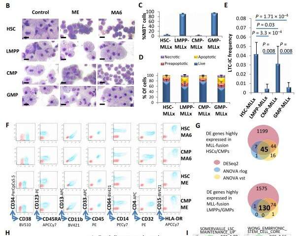
图1.人HSC和CMPS但不是LMPPS或GMP是MLL融合介导的转化的细胞靶标。(a)每周每周测量培养细胞的细胞的增殖,用于正常和MLL转化的细胞种群。我,mll-ild;MA6,MLL-AF6。(b)在4至6周的典型细胞形态在4至6周的表明细胞类型的体外培养。秤条,10m。(c)NBT染色定义指定基团的髓样区分化细胞的百分比(每种胞胎n = 4)。mllx:mll-ild和mll-af6。P = 1.25×10⁻¹⁹,HSC-/ CMP-MLLX与LMPP-/ GMP-MLLX。(D)条形图代表膜蛋白V和指定细胞的培养纤维术后每类别的细胞百分比。 Data are presented as means ± SD, and statistical significance of P = 0.00319 was determined by ANOVA from N=3, n=6, F- value=14.86, df=1, for HSC-/CMP-MLLx versus LMPP-/GMP-MLLx. (E) Longterm culture initiating cell (LTC-IC) frequencies of the indicated groups at 4 weeks of in vitro culture (n = 6). (F) Typical surface marker abundance of the indicated transformed cell populations at 8 to 10 weeks in culture. (G) Venn diagram showing the overlap of significantly differentially expressed (DE) genes between HSC/CMP-MLLx and LMPP/ GMP-MLLx cells using DESeq2, ANOVA (on rlog transformed read counts) and ANOVA (on vst transformed read counts) as indicated. (H) Heatmap showing the 130 genes with consistently higher expression in LMPP/GMP-MLLx and the 45 genes with consistently higher expression in HSC/CMP-MLLx and their functional annotations according to webgestalt and Toppgene. (I) Selected stem cell–related gene sets from GSEA are shown comparing HSC-/CMP-MLLx (HC-MLLx) and LMPP-/GMP-MLLx (LG/MLLx) cells. In (A), (C), and (E), data are represented as means ± SD. In (C) and (E), statistical significance was determined by Student t test. Credit: Zeisig et al., Sci. Transl. Med. 13, eabc4822 (2021) 24 February 2021


用户评论