一个决定与组织水平和可伸缩的封装材料杨氏模量。)示意图bioelectronic接口的周围神经和软导体电极和绝缘材料。b)逐步PEDOT的示意图:PSS决定合成工艺和SEM图像显示形态变化在每一步合成的一个决定。c)特区电导和电导率变化在过渡从离子凝胶电解珩磨泡在水里。d)在转换过程中体积变化从离子凝胶电解珩磨。f)电化学阻抗的变化在不同的单轴压力在1赫兹,100赫兹和1 kHz电场频率。决定膜厚度在应变0% 200海里。y g,表面阻抗在不同压力和频率的阴谋。h)单轴应力-应变曲线的大部分决定样本。i)的分子结构弹性PFPE-DMA (dimethacrylate-functionalized perfluoropolyether)进行交联过程后暴露于紫外线光。 j) Uniaxial stress–strain curve of the crosslinked fluorinated elastic PFPE-DMA. k) Comparison of Young’s modulus values between commonly used dielectric materials and the conductor with PFPE-DMA and ECH. Conductive and insulation materials are shaded in pink and blue, respectively. Credit:自然生物医学工程,doi: https://doi.org/10.1038/s41551 - 018 - 0335 - 6
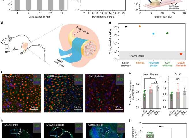
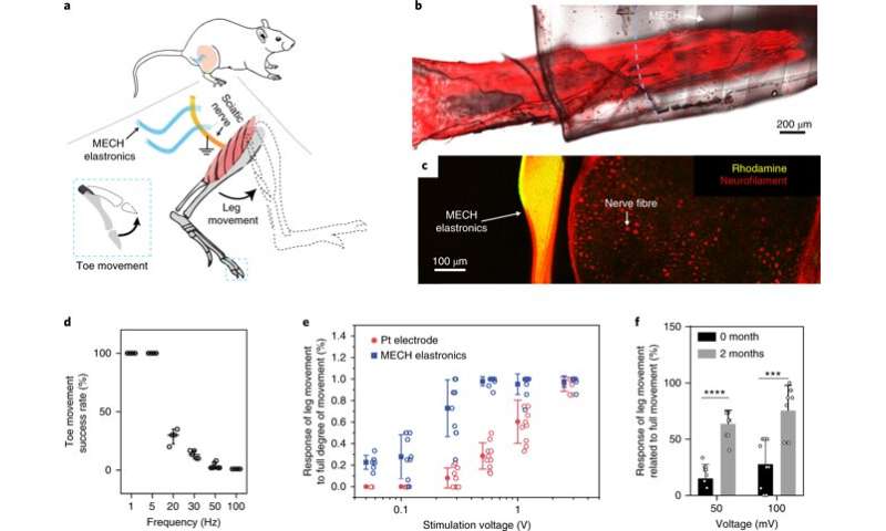
小说的电极阵列薄膜水凝胶被称为“elastronics。”They are 20 µm in feature size, with significantly reduced interfacial impedance in the surrounding tissue. The system contains a current-injection density approximately 30 times greater than platinum electrodes and shows stable electrical performance under strain. The scientists demonstrated the use of soft elastronic arrays for localized, low-voltage electrical stimulation of the sciatic nerve in live mice.
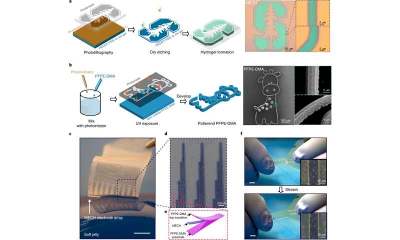
光刻水凝胶elastronics。左一):逐步插图的光刻装置。传统光刻进行PEDOT: PSS-ionic液体(IL)离子凝胶盟硬掩模。之后,微型图象被干蚀刻转移到离子凝胶。最后,微型图象离子凝胶被水交换了。右:复杂的微观结构装置,直线和曲线都解决了。b)左:逐步说明对光刻PFPE缩微成像。PFPE-DMA混合剂和旋转涂布。紫外线是用来交联PFPE-DMA形式感兴趣的微型图象。右:扫描电镜的图像photolithographically微型图象PFPE-DMA结构直线和曲线。 c) A freestanding MECH elastronics electrode array pressed against soft jelly. Scale bar, 2 mm. d) Zoomed-in image of MECH electrodes (dark lines) with PFPE-DMA encapsulation (colored as light blue). e) Schematic of an elastronic electrode, a MECH electrode and interconnect sandwiched by photolithographically micropatterned fluorinated polymer PFPE-DMA as the top and bottom insulation layers. f) MECH electrode array stretched under 20% tensile strain shows no cracks. Credit: Nature Biomedical Engineering, doi: https://doi.org/10.1038/s41551-018-0335-6
 Schematic of the bioelectronic interface between a peripheral nerve and soft conductor electrodes and insulation materials. b) Schematic of the stepwise PEDOT:PSS ECH synthesis process and SEM images showing morphological changes in each step during the synthesis of an ECH. c) d.c. conductance and conductivity change during the transition from ion gel to ECH by soaking in water over time. d) Volume change during the transition from ion gel to ECH. f) Change in electrochemical impedance under different uniaxial strains at 1 Hz, 100 Hz and 1 kHz electrical field frequency. ECH film thickness is 200 nm at 0% strain. y. g, Surface plot of impedance at different strains and frequencies. h) Uniaxial stress–strain curve of the bulk ECH samples. i) Molecular structure of elastic PFPE-DMA (dimethacrylate-functionalized perfluoropolyether) undergoing the crosslinking process following exposure to UV light. j) Uniaxial stress–strain curve of the crosslinked fluorinated elastic PFPE-DMA. k) Comparison of Young’s modulus values between commonly used dielectric materials and the conductor with PFPE-DMA and ECH. Conductive and insulation materials are shaded in pink and blue, respectively. Credit: <i>Nature Biomedical Engineering</i>, doi: https://doi.org/10.1038/s41551-018-0335-6")


用户评论